
 English
 Spanish
 Turkish
Functions and Precautions of Otoscope
Functions and Precautions of Otoscope
An otoscope is a medical device used to examine the ear canal and eardrum. It consists of a slender tube, a high-resolution camera, and a light source, providing clear, magnified real-time images of the ear. This assists doctors in accurately diagnosing ear conditions such as otitis media, foreign objects in the ear canal, or eardrum perforations. The procedure is non-invasive, quick, and commonly used in otology.
1. Definition and Functions of Otoscope
The otoscope projects the internal structures of the ear canal onto a display screen via a front-mounted miniature camera, enabling visual examination. Its core functions include:
Detailed Observation:
Magnifying the surface of the ear canal and eardrum to detect lesions difficult to see with the naked eye (e.g., microperforations, bleeding spots).Assisting Diagnosis:
Identifying the causes of symptoms such as ear pain, hearing loss, or ear discharge.Guiding Treatment:
Facilitating procedures like removal of foreign objects or earwax (cerumen), or performing minimally invasive operations such as eardrum repair.
2. Examination Process and Applicable Scenarios
Examination Steps:
The patient sits while the doctor adjusts the light and lens angle.
The otoscope tube is gently inserted to avoid touching sensitive areas.
The ear canal and eardrum are observed via the display screen, recording the location and morphology of any lesions.
Applicable Situations:
Recurrent ear pain, ear fullness, or tinnitus.
Suspected foreign objects or cerumen impaction in the ear canal.
Post-trauma assessment of eardrum integrity.
Post-operative follow-up or disease progression monitoring.
3. Precautions and Limitations
Operational Requirements:
Must be performed by a professional doctor to avoid ear canal injury due to improper operation.Contraindications:
Examination should be postponed in cases of acute ear canal infection, severe swelling, or bleeding.Limitations:
Unable to penetrate bone or soft tissue; conditions affecting the middle or inner ear require additional tests such as CT scans or hearing assessments.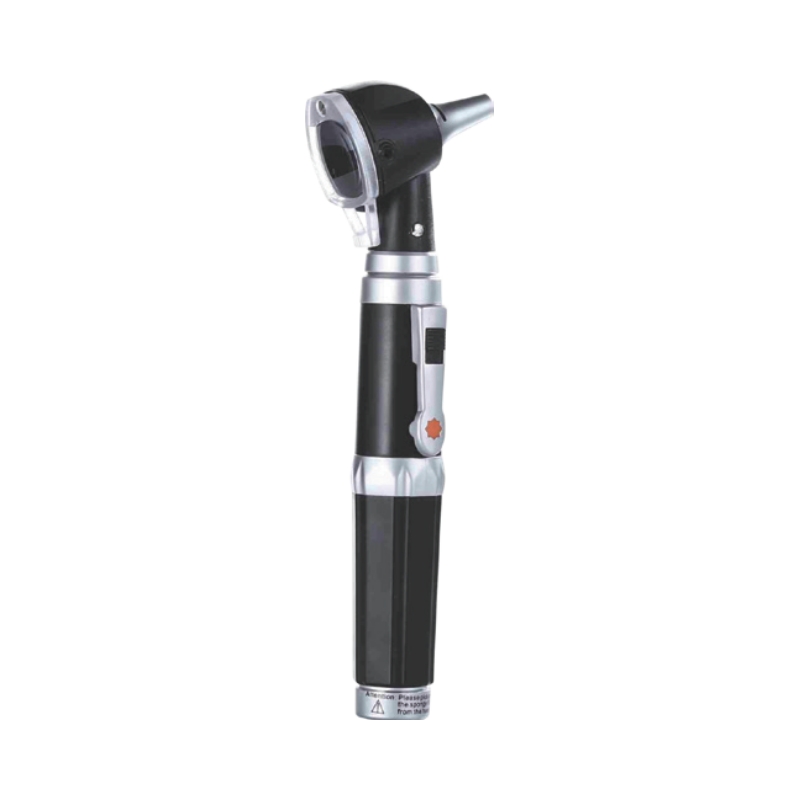
4. Technical Advantages Compared to Traditional Methods
Compared to traditional methods like frontal mirrors or conventional otoscopes, the modern otoscope offers:
High-Definition Imaging:
Reduces misdiagnosis rates, especially suitable for children or individuals with narrow ear canals.Dynamic Recording:
Supports image storage for pre- and post-treatment comparison.Patient Involvement:
Patients can visually understand their condition via the screen, improving doctor-patient communication.
5. Conclusion
The otoscope is a vital tool in diagnosing ear diseases, enhancing examination accuracy through visualization. However, results should be interpreted in combination with patient symptoms and other tests. If ear discomfort occurs, seek medical attention promptly. Avoid self-operating devices or digging the ear canal to prevent further injury.


