
 English
 Spanish
 Turkish
The Essential Role of Fundus Cameras in Modern Eye Care
The Essential Role of Fundus Cameras in Modern Eye Care
A fundus camera represents a critical diagnostic tool in ophthalmology and optometry, enabling the detailed photographic documentation of the interior surface of the eye. This includes the retina, optic disc, macula, and retinal vasculature. The primary purpose of a fundus camera is to capture high-resolution images for the detection, monitoring, and management of ocular diseases such as diabetic retinopathy, glaucoma, macular degeneration, and hypertensive retinopathy. By providing a permanent, objective record of the retina's condition, a fundus camera facilitates accurate diagnosis, tracks disease progression over time, and enhances communication between eye care professionals and their patients. The integration of a reliable fundus camera into a practice is fundamental to delivering comprehensive eye health services.
Understanding How a Fundus Camera Works
To appreciate its clinical value, one must understand the basic operation of a fundus camera. This specialized instrument functions by projecting a beam of light through the pupil to illuminate the retina. A precisely aligned internal camera then captures the reflected light, creating a detailed two-dimensional image of the intricate retinal structures. Modern digital fundus cameras have largely replaced film-based systems, allowing for instant image review, digital storage, and easy sharing. The sophistication of a fundus camera lies in its optical system, which must provide sufficient illumination while managing reflections and offering a wide enough field of view to capture clinically significant areas of the retina.
Key Applications and Clinical Settings for Fundus Imaging
The utility of a fundus camera spans numerous clinical scenarios, making it indispensable in various settings. In standard ophthalmic and optometric practices, it is routinely used for annual diabetic eye screenings, which are crucial for preventing vision loss. Glaucoma specialists rely on fundus camera images to assess and monitor the optic nerve head for signs of damage. Retinal specialists utilize these images to diagnose and manage conditions like age-related macular degeneration (AMD) and retinal detachments. Beyond specialized clinics, a portable fundus camera is increasingly valuable in primary care facilities, community health screening programs, nursing homes, and even corporate wellness checks, helping to bridge gaps in access to essential eye care.
Advantages of Non-Mydriatic Fundus Cameras in Patient Care
A significant advancement in retinal imaging is the non-mydriatic fundus camera. Traditional cameras often require pupil dilation using eye drops, a process that can be uncomfortable for patients, causing temporary blurred vision and light sensitivity. The non-mydriatic fundus camera is designed to capture high-quality images through an undilated pupil, typically using infrared light for alignment and a low-power flash for capture. This technology greatly enhances patient comfort, improves workflow efficiency by eliminating the 15-20 minute dilation wait time, and increases the feasibility of large-scale screening programs. For many patients and busy clinics, a non-mydriatic fundus camera is the preferred choice for accessible and efficient retinal documentation.
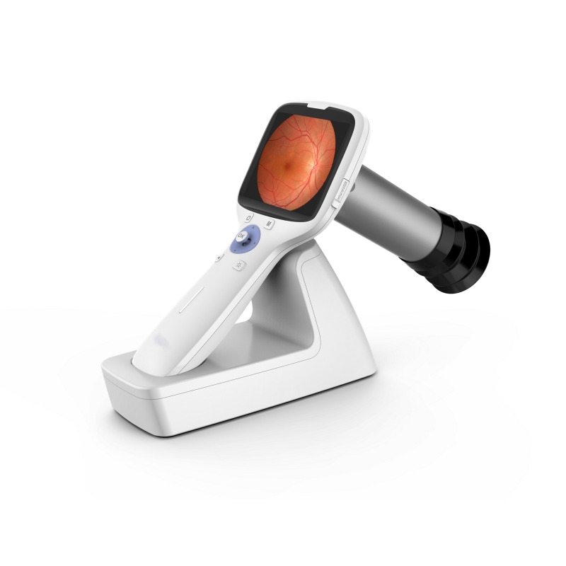
Introducing the KELLYUNION Portable Non-Mydriatic Fundus Camera
The KELLYUNION Fundus Camera embodies the evolution of retinal imaging technology, combining diagnostic power with exceptional convenience. Designed with the modern practitioner in mind, this device addresses the core needs of accuracy, efficiency, and mobility in patient care. As a non-mydriatic fundus camera with a 45° viewing angle, it provides a balanced field of view sufficient for capturing key retinal landmarks without requiring patient dilation. This feature alone makes the KELLYUNION fundus camera a versatile tool for rapid assessment and screening in diverse environments, from the main clinic to outreach locations.
Core Features and Technical Specifications of the Device
The KELLYUNION Fundus Camera is engineered to deliver outstanding performance through a suite of user-focused features. Its core strength lies in acquiring stable, high-quality retinal images that support confident clinical decisions. The device integrates essential functions including photography, image saving, and real-time observation on its display, streamlining the examination process. A defining characteristic of this fundus camera is its integrated WiFi transmission capability. This allows captured images to be instantly and securely transmitted to a computer, tablet, or other mobile devices, facilitating easy integration into electronic health records (EHR), remote consultation, or immediate patient education.
Unmatched Portability for Outreach and Flexible Diagnostics
Beyond its imaging capabilities, the KELLYUNION fundus camera is distinguished by its portable design. Lightweight and easy to carry, this fundus camera is specifically built to meet the demands of mobile diagnostics and doctors' requirements for outpatient or community visits. Its portability ensures that high-standard retinal imaging is no longer confined to a fixed examination room. Healthcare providers can now effortlessly bring this fundus camera to satellite clinics, health fairs, nursing homes, or home visits, dramatically expanding the reach of vital preventive eye care services and making the device an invaluable asset for any outreach initiative.
Enhancing Clinical Workflow and Patient Management
Implementing the KELLYUNION Fundus Camera into a practice significantly enhances operational workflow and patient management. The immediacy of digital images allows for on-the-spot review and analysis with the patient, fostering better understanding and engagement in their eye health. The combination of non-mydriatic operation and efficient workflow reduces overall appointment times, enabling clinicians to see more patients without compromising care quality. Furthermore, the digital records created by this fundus camera simplify the tracking of retinal changes over years, providing a clear, visual history that is crucial for managing chronic ocular conditions effectively and demonstrating the necessity of treatment plans to patients.
A Commitment to Quality with KELLYUNION
KELLYUNION stands as a trusted global partner in the medical device industry, with a comprehensive portfolio of health technology solutions. Our commitment is unwavering: to supply medical professionals worldwide with safe, reliable, and effective equipment. This commitment is underscored by our adherence to rigorous international quality standards and holding relevant certifications for our devices. Our extensive distribution network supports clients across the globe, ensuring that innovative and essential tools like the KELLYUNION Fundus Camera are accessible wherever quality eye care is provided.
Discover how the portable KELLYUNION Non-Mydriatic Fundus Camera can transform retinal screening in your practice. For detailed specifications and to explore our full range of ophthalmic solutions, visit our official website at www.kellyunion.com.


